HO-200 Ophthalmic Ultrasound Scanner Chinese manufacturer
Healicom Medical is a Chinese manufacturer specializing in ophthalmic ultrasound equipment. We are committed to providing high-performance ophthalmic ultrasound equipment to meet your needs. With decades of combined experience in the medical device industry, we have advanced manufacturing facilities and have accumulated extensive experience in ophthalmic ultrasound projects.
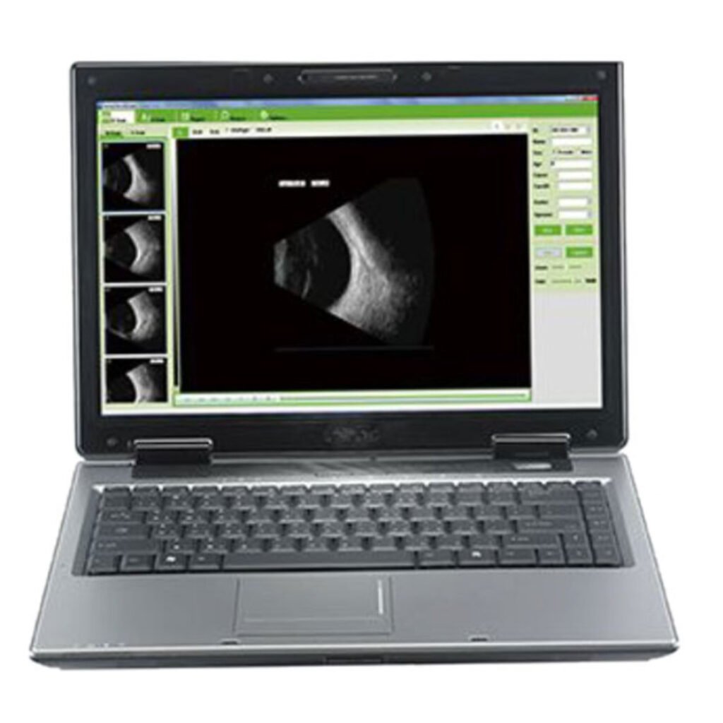
HO-200 A/B Scan with normal, vitreous body enhancement, retina observation mode, mainly used for diagnosis of intraocular diseases, display the location, shape range of the focus of infection and the relationship with the surrounding tissue. Can be diagnosed vitreous opacity, retinal detachment, eye base tumors etc. eye diseases. A scan is used to measure anterior chamber depth, lens thickness, axial length, calculate diopter of implant IOL as well.
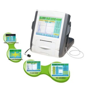
A-scan parameters include 10MHz frequency, equipped with LED, depth of 40mm, and accuracy of ±0.05mm. Measurements included anterior chamber depth, lens thickness, vitreous length, total length, and average. Supports different eye modes such as phakic, aphakic, intensive and various intraocular lens (IOL) modes. IOL formulas include SRK-II, SRK-T, HOFFER-Q, HOLLADAY, BINKHORST-II, and HAIGIS. Calculation functions include mean and standard deviation, with the ability to store 10 scans for each eye.
B-scan parameters include 10MHz and 20MHz (optional) frequencies, using magnetic drive technology for noise-free operation. The scanning mode is sector scanning, with multi-level continuous amplification and real-time amplification functions. The resolution is ≤0.3mm in the lateral direction and ≤0.2mm in the longitudinal direction. The geometric position accuracy is ≤10% in the lateral direction and ≤5% in the longitudinal direction. The scanning depth is 60mm, the vitreous and retinal parts are enhanced, and the probe gain range is 30dB to 105dB. 53° scanning angle and 256 levels of grayscale, and supports a variety of pseudo-color displays. Measurement types cover multiple sets of distances, perimeters, and areas. Image post-processing functions include multi-curve processing and pseudo-color processing curves. Supports movie playback of 100 frames of images, and the output formats are AVI and JPG.
Others
Other parameters include display modes: B, B+B, B+A, A mode, which can prompt and search based on preset keywords, and support multi-keyword search cases. The device runs on Windows XP, Vista and Windows 7 platforms, ensuring broad compatibility and ease of operation.
Application Scenario Analysis
Ophthalmic ultrasound technology is widely used in various ophthalmic clinical and diagnostic scenarios. The following is an analysis of some major application scenarios:
Pre-cataract surgery evaluation: A-scan: used to measure the axial length of the eye, the depth of the anterior chamber and the thickness of the lens to accurately calculate the power of the intraocular lens (IOL). Commonly used formulas such as SRK-II, SRK-T, HOFFER-Q, HOLLADAY, etc. can help doctors choose the appropriate IOL type.
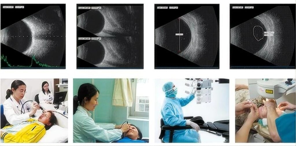
Diagnosis of retinal and vitreous diseases: B-ultrasound scan: Through sector scanning mode and high-resolution imaging, it can clearly display retinal detachment, vitreous hemorrhage, vitreous opacity and other lesions. Enhancing the function of parts of the vitreous and retina can help doctors perform detailed localization and assessment of lesions.
Eye tumor detection: B-ultrasound scan: High-resolution imaging technology can be used to detect the location, size and nature of tumors within the eye. Multiple pseudo-color displays and multi-curve processing functions help doctors better identify and analyze lesions.
Glaucoma Diagnosis and Management: A-scan: used to measure anterior chamber depth to help diagnose and manage glaucoma. Accurate measurement of anterior chamber depth is critical for assessing angle status and determining treatment options.
Eye injury assessment: B-ultrasound scan: In the case of eye trauma, the scan can evaluate the integrity of the internal structures of the eyeball, including the condition of the lens, vitreous body, and retina. Rapid, accurate diagnosis can provide important information for emergency treatment.
Examination of children and uncooperative patients: B-ultrasound scan: For patients who are difficult to cooperate, such as children or patients with intellectual disabilities, B-ultrasound can provide clear intraocular images without excessive cooperation of the eyes, making it convenient for doctors to conduct examination and diagnosis.
Comprehensive evaluation of complex eye diseases: Comprehensive display mode (B, B+B, B+A, A): Different display modes can be selected according to specific needs to provide more comprehensive eye structure information and help doctors conduct comprehensive assessment and diagnosis of complex eye diseases.
These application scenarios demonstrate the importance and widespread application of ophthalmic ultrasound in modern ophthalmic diagnosis and treatment. Through precise measurement and high-resolution imaging technology, ophthalmic ultrasound provides doctors with a powerful diagnostic tool, helping to improve the effectiveness of diagnosis and treatment and Patient satisfaction.
Request A Free Quote
You can send us your enquiry and we will reply you in time.
We’d like to work with you
Send us a message if you have any questions or request a quote. Our experts will give you a reply within 24 hours and help you select the right product you want.