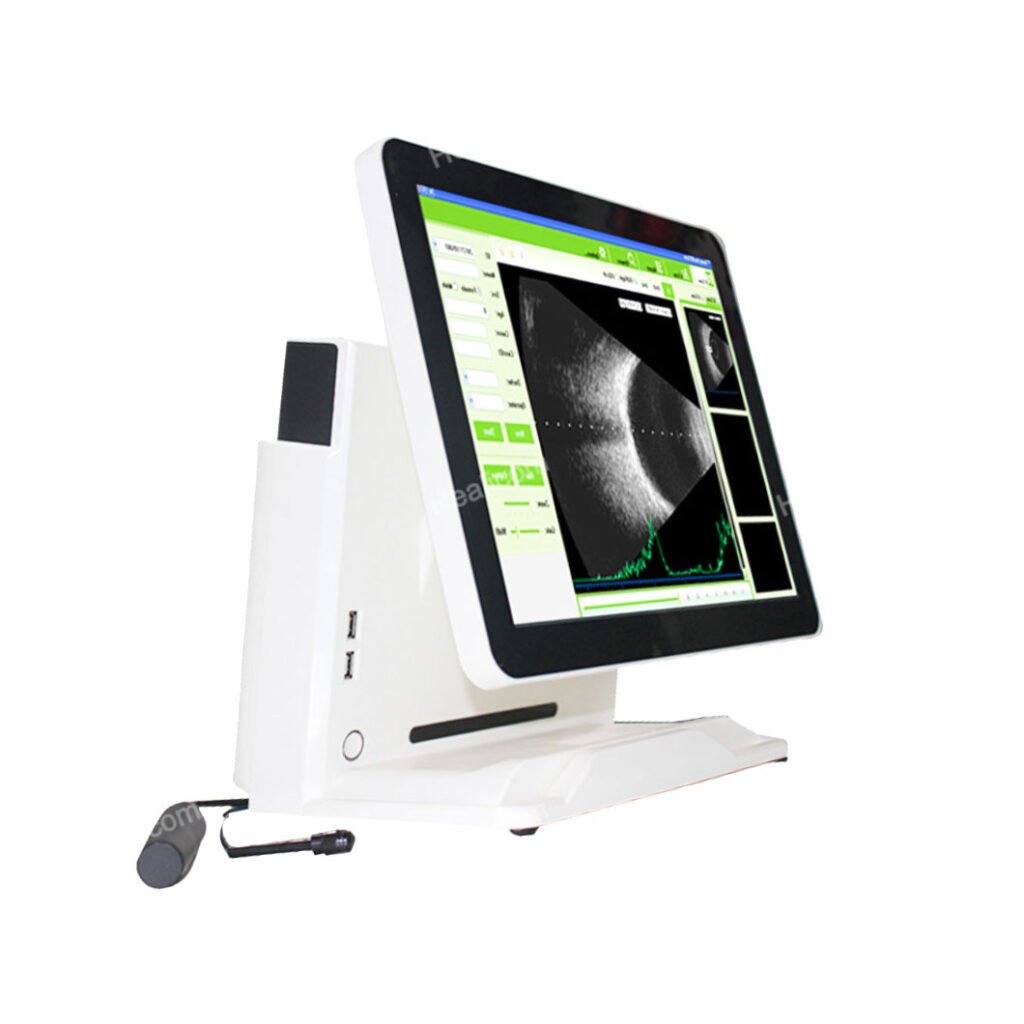
HO-500 A/B Scan with normal, vitreous body enhancement, retina observation mode, mainly used for diagnosis of intraocular diseases, display the location, shape range of the focus of infection and the relationship with the surrounding tissue. Can be diagnosed vitreous opacity, retinal detachment, eye base tumors etc. eye diseases. A scan is used to measure anterior chamber depth, lens thickness, axial length, calculate diopter of implant IOL as well.
An Ultrasound Ophthalmic A/B Scanner is a medical device used to visualize eye structures, diagnose conditions, and guide treatments, providing detailed A-scan and B-scan ultrasound images.
-15 inch LED touch screen, all-in-one, pretty, portable
-Integrated Image Capture
-Integrated Patient Database
-Integrated Report Editor
-Can work with battery
Product Name | Ophthalmic A / B Ultrasound Scanner |
Model | HO-500 |
B scan | |
Frequency | 10MHz/20MHz (optional) ,Magnetic driven, noiseless |
Scanning Mode | Sector Scanning |
Magnify | Multi continuous magnification,Real-Time magnification |
Resolution | Lateral ≤0.3mm; Vertical≤0.2mm |
Geometry position precision | Lateral ≤10%; Vertical≤5% |
Depth | 60mm |
Enhance the part of vitreous body and retina | |
Gain of probe | 30dB-105dB |
Scanning Angle | 53° |
Gray Scale | 256 |
False Color | Multi colors. OCT |
Measurement type | Multi group distances, perimeters and areas |
Image post processing | multiple curves processing, Pseudo-color processing curve |
Movies | 100 images movie review, AVI JPG format image output |
A scan | |
Frequency | 10MHz, with LED |
Depth | 40mm |
Precision | ±0.05 mm |
Measurement | Anterior chamber depth, lens thickness, vitreous body length, total length and average |
Eye mode | Phakic / Aphakic / Dense / Various |
IOL IOL Formula | SRK-II, SRK-T, HOFFER-Q, HOLLADAY, BINKHORST-II, HAIGIS |
Stat. Calculation | Average and standard deviation |
Store | 10 Scanning results for each eye |
Others | |
Display Mode | B, B+B, B+A, A |
Hint | preset keyword |
Case Search | Multi-keywords |
Screen | 15 inch LCD |
Built-in battery | can use 4 hours |
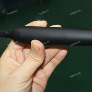
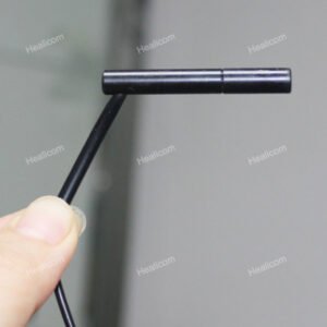
Product detail drawings help users understand the design and functional layout of the product by showing the appearance and internal structure of the device. It shows the location of key components and interfaces to facilitate installation and operation, and provides specification and dimensional information to assist purchasing decisions and space planning. In addition, detailed drawings help users identify brand logos and model numbers to ensure product authenticity and compatibility.
· Diagnosis of intraocular and periocular diseases, such as retinal detachment, vitreous hemorrhage, tumors, etc.
· Measure axial length and corneal thickness for preoperative evaluation such as cataract surgery.
· Assess structural changes within the eyeball, especially in the presence of opaque media such as cataracts or vitreous opacities. Monitor the progression of glaucoma, intraocular inflammation, and other chronic eye diseases.
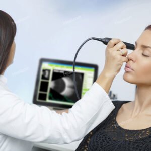
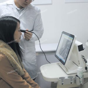
· Professional medical personnel perform the operation and interpret the scan results.
· Ensure proper calibration and sterilization of equipment to prevent cross-contamination.
· Select the appropriate scanning mode and parameters according to the patient’s condition.
· For special groups, such as children, pregnant women or patients with special medical conditions, caution should be used.
· Pay attention to the impact of ultrasound on eye tissue and avoid over-scanning and damage.
· Regularly maintain and calibrate equipment to ensure its performance and accuracy.
You can send us your enquiry and we will reply you in time.
Send us a message if you have any questions or request a quote. Our experts will give you a reply within 24 hours and help you select the right product you want.
Get all the latest news, exclusive deals and offers.